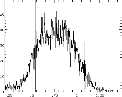
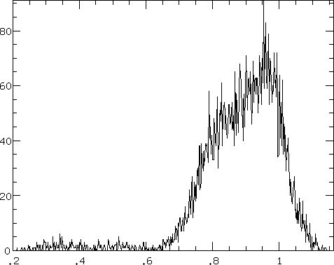
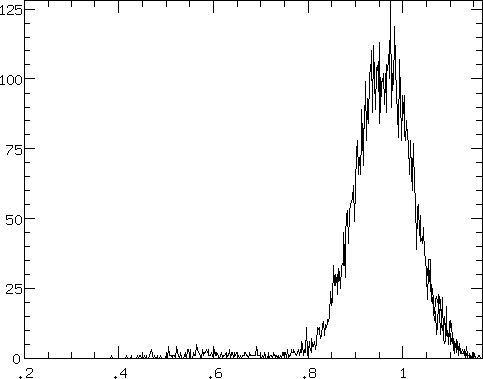
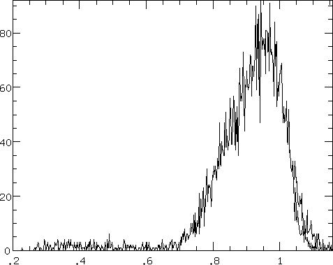
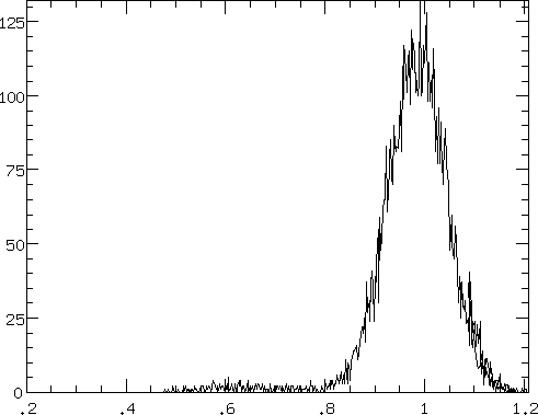
The plots on this page are histograms of the images created by
dividing two flat field images of the same energy. In the first
column, the (pre-fix) flat field image using the XRCF HV and
XRCF threshold
voltage settings is divided by the post-fix flat field, which used the
flight level HV and flight threshold voltages. In the second column,
the (pre-fix) flat field image which used the flight HV but the XRCF
threshold voltage are again divided by the post-fix flat field.
All of the histograms are based on the upper 65% of all the images,
in order to avoid the regions affected by vignetting. All of the images
were blocked with a blocking factor of 128.
The ratio images have all been normalized by dividing the entire
image by the image cell average in a 121-cell central region.
The histogram profiles therefore provide a measure of the relative
flatness of the images taken at differing voltage settings.
Note that the first column profiles are consistently broader and
more highly skewed than those of the second column. The ratio
of profile widths (excluding outliers) for each energy are:
| Oxygen | 2.3 |
|
| Aluminum | 1.5 |
| Titanium | 1.5 |
These graphs indicate the degree to which the change in detector HV affects
detector response. The response is much
more greatly affected by the change in the HV setting than the change
in threshold voltage.
Boron: XRCF HV over post-fix flight settings
 |
Boron: flight HV over post-fix flight settings
The pre-fix flight HV data are
missing. |
Oxygen: XRCF HV over post-fix flight settings
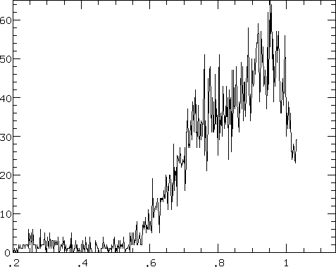 |
Oxygen: flight HV over post-fix flight settings
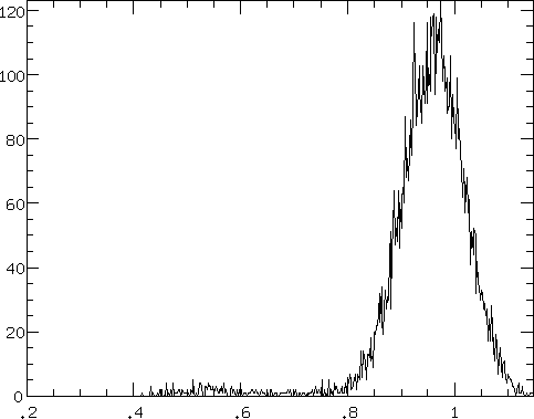 |
Aluminum: XRCF HV over post-fix flight settings
 |
Aluminum: flight HV over post-fix flight settings
 |
Titanium: XRCF HV over post-fix flight settings
 |
Titanium: flight HV over post-fix flight settings
 |